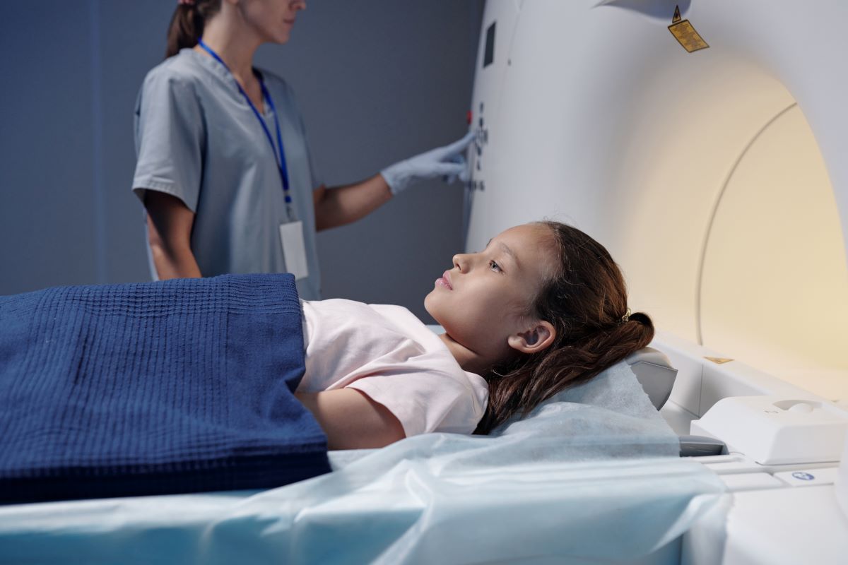
A recent study featuring research by University of Florida Engineering Professor Wesley Bolch, Ph.D., links exposure to radiation from medical imaging to a small-but-significant risk of blood cancers among children and adolescents.
But do not panic. The study concludes the benefits of medical imaging outweigh the minimal risks.
Funded by the National Cancer Institute, the study will help medical personnel make informed decisions about using imaging on children. The study concluded that while ionizing radiation is a carcinogen, the benefit-to-risk ratio favors CT imaging of children when imaging is justified and the technique minimizes adverse effects.
“These risks are low and when justified by the imaging physician, patient benefits, such as disease detection, will greatly outweigh these very small risks.” —Wesley Bolch, Ph.D., University of Florida engineering professor
The paper, “Medical Imaging and Pediatric and Adolescent Hematologic Cancer Risk,” was published last week in the New England Journal of Medicine. For his part of the study, Bolch used virtual patient anatomic models to reconstruct bone marrow doses in more than 3.7 million children who underwent CT imaging between 1996 to 2016.

“We used a library of 3D anatomic whole-body computerized patient models developed in the early 2010s under a contract with the National Cancer Institute,” said Bolch, a distinguished professor in biomedical and radiological engineering with UF’s J. Crayton Pruitt Family Department of Biomedical Engineering.
In Bolch’s Advanced Laboratory for Radiation Dosimetry Studies, researchers developed hundreds of models representative of U.S. adults and children across all combinations of ages, heights and weights.
“We call this an organ dose reconstruction,” said Bolch, who is also a member of the UF Health Cancer Center.
The study is unique because it directly measures leukemia risks in these pediatric patients. Previously, researchers used established cancer-risk models, many of which were based on data from the atomic bomb survivors in 1945 Japan. Needless to say, Bolch noted, diagnostic X-ray exposures are vastly different from atomic weapon radiation fields.
“This is the very first study of its kind in the U.S. and Canada, and the very first study of cancer risks in children undergoing medical imaging where each patient was considered in a unique manner regarding their sex, body size and medical imaging exposure technique factors,” he said.
While CT imaging contributes a greater fraction of total radiation, doses from nuclear medicine, radiography and fluoroscopy were also included for bone marrow-dose calculations.
“Dr. Bolch’s role in this landmark study highlights UF BME’s leadership in making medical imaging safer for children. It demonstrates UF’s dedication to research that safeguards patients and informs global healthcare practices,” said BME chair Cherie Stabler, Ph.D.
In the study, the highest CT doses to bone marrow were seen in head-and-neck imaging, where the average dose was 30.8 milligray (a unit measuring ionizing radiation deposited in tissue). CT imaging of the head – one of the more common pediatric CT scans – showed an average dose of 13.7 milligray.
The incidence of hematologic cancers by age 21 years was 0.3% among those children exposed to bone marrow doses more than 30 milligray. However, fewer than 1% of the 3.7 million children in this study had cumulative doses exceeding 30 milligray.
Plus, CT imaging doses today are much lower, and imaging systems are much faster than in the late-1990s and early-2000s.
This research comes 25 years after a Columbia University research paper made the link between leukemia and some radiology scans, thus scaring “every mother in this country,” Bolch said.
The key issue in that study showed imaging technologists and radiologists were not making adjustments to X-ray techniques that explicitly considered the size of the patient.