Manager of Core and Laboratory Infrastructure
(352) 273-7231
KHines@bme.ufl.edu
The equipment described below is shared instrumentation available to primary UF BME faculty members available in the Biomedical Sciences Building:
IMAGING
Zeiss LSM 880 Confocal Microscope with airy scan
Located in BMS J320
This device provides high resolution, sensitivity and speed that increases productivity when performing quantitative imaging. Its unique Airyscan detector enables super resolution imaging (120 nm resolution in XY, 400 nm in Z). It includes laser lines 405, 458, 488, 514, 561, 633 nm. Our system also includes a full incubation system for live cell imaging using heat and CO2.
Zeiss LSM710 Confocal Microscope
Located in BMS J238A
This high precision confocal microscope comes with 5 lasers (405, 488, 514, 546, 688 nm) and offers great sensitivity and less noise, due to longer sampling time per pixel and excellent contrast characteristics
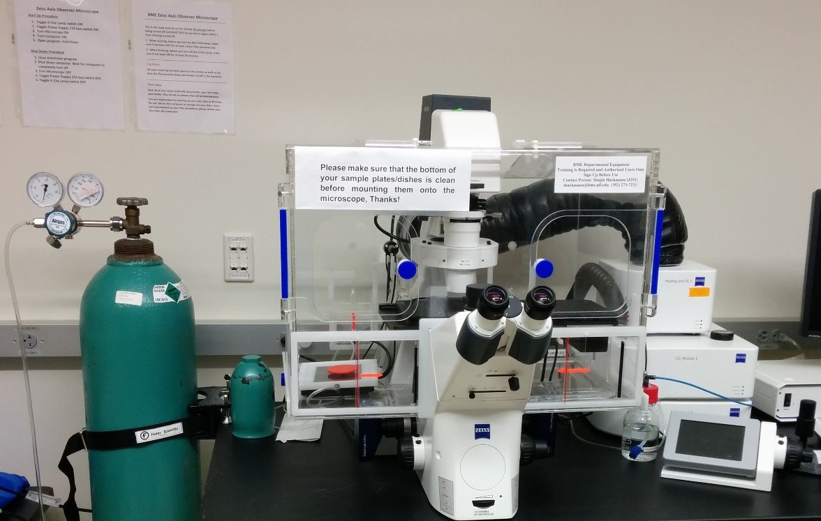
Zeiss High Resolution Inverted Fluorescence Microscope
Located in BMS J238A
This device offers bright field, dark field, phase contrast, differential interference contrast and fluorescence (FITC, Cy3, Cy5 and DAPI) and provides sharp image contrasts for clear imaging. Our system also includes a full incubation system for live cell imaging using heat and CO2
Perkin Elmer IVIS Spectrum CT In Vivo Imaging System
Located in BMS J320
This is an in vivo imaging system to facilitate non-invasive longitudinal monitoring of disease progression, cell trafficking and gene expression patterns in live animals. It also offers single-view 3D tomography for both fluorescent and bioluminescent reporters that can be analyzed in an anatomical context. It offers High resolution (to 20 microns) with 3.9 cm field of view, with twenty eight high efficiency filters spanning 430 – 850 nm. This instrument can do 3D diffuse tomographic reconstruction for both fluorescence and bioluminescence.
Momentum Magnetic Particle Imaging (MPI)
Located in BMS J238A
Magnetic Particle Imaging (MPI) is a molecular imaging modality that measure the location and concentration of superparamagnetic nanoparticle tracers in vivo (typically Iron Oxide, SPIOs) by detecting their response to spatially dependent magnetic fields.
LI-COR Odyssey CLx Imaging Station
Located in BMS J228
This is an infrared imager features two spectrally separate solid state lasers (685 nm and 785 nm) to detect infrared dye-labeled conjugates and near-infrared stains. This device generates imaging results in the much cleaner background, higher signal-to-noise ratios and better sensitivity, as compared to standard imaging system.
HISTOLOGY CORE
Location is BMS J217 for all equipment other than the embedding station
General Data ETC 2120 Histology Embedding Station
This equipment embed tissue samples and create paraffin embedded tissue blocks.
The Embedding Station is located in BMS J220
Leica Rotary Microtome RM2125 RTS
This manually operated device is used for creating thin sections of specimens of varying hardness. Is designed for sectioning soft paraffin specimens. We have two of microtomes available for use.
Optical Inverted microscope – A light source driven optical microscope is available for use in the Histology core, while working on sectioning.
Leica CryoStat CM1950
This is a highly adaptable device used to do frozen section histology at a temperature as low as -20 degrees C
Chemical Fume Hood – A second fume hood is available for additional work in the BMS J217 Histology Core.
VWR Water Bath – This is an unstirred water bath that can hold a constant temperature. We have two water baths available for use.
CELL & MATERIALS CHARACTERIZATION
Thermofisher QuantStudio Real-Time qPCR
Located in BMS J222
The system supports a broad range of real-time genomic applications, such as analyses of gene expression, microRNAs and noncoding RNAs, copy number variation, drug metabolism enzymes, and protein expression; SNP genotyping; and mutation detection
BD FACS Celesta Flow Cytometer
Located in BMS J228
This is a multicolor flow cytometer that comes with 3 lasers (blue, red and violet) and can be employed in cell counting and biomarker detection.
Molecular Devices M5E plate reader
Located in BMS J223
This is a temperature controlled (4°C above ambient to 50°C) plate reader that can measure fluorescence, luminescence, absorbance, time-resolved fluorescence (TRF) and fluorescence polarization (TRP). It has dual monochromators that allows users to determine optimal excitation and emission settings for both fluorescence and absorbance. This instrument can handle 6-, 96-, and 384- well plates and allows for end point, kinetic, spectral scanning, and well-area scanning modes.
BASIC LABORATORY EQUIPMENT
Sorvall RC 6 Plus Superspeed Centrifuge
Located in BMS J243
This high speed, high capacity, refrigerated centrifuge offers 4 liters of capacity and processing speeds of up to 22,000 rpm enabling fast processing of samples
Sorvall LYNX 6000 Superspeed Centrifuge
Located in NEB 389
This high speed, high capacity, refrigerated centrifuge offers 6 liters of capacity and processing speeds of up to 29,000 rpm enabling fast processing of samples. It is the newer version of the RC 6, and is located in New Engineering Building
Sorvall WX Ultra 90 Floor Ultra Centrifuge
Located in BMS J361
This high performance, refrigerated ultra centrifuge will allow nanoparticle separation at up to 90,000 rpm.
Refrigerator and -4 degree freezer
Located in NEB 389
For temporary overflow storage, please contact Kelley Hines with description of needs
Chest freezer
Located in NEB 383
For temporary overflow storage, please contact Kelley Hines with description of needs
Large Biosafety Hood
Located in NEB 389
8 foot calibrated hood available for use in NEB 389
Laboratory Ice Machines
Located in NEB 383, and 2nd and 3rd floor back hallways of BMS
Serviced, filtered ice machines for laboratory ice needs only